Banana cells
Compound microscope activity sheet
Materials
- Pasteur pipette
- Unripe banana
- Microscope slides
- Cover slips
- Paper towels
- Lugol Solution
- Tooth pick
Methods
- Smear a little (less than the size of a sesame seed) of an unripe green banana on a microscope slide to rub the cells apart.
- Place a drop of Lugol solution on top of the banana smear.
- Place a coverslip on top and remove the excess of solution with a tissue.
- Place the slide on the microscope, with 4 x or 10x objective in position and find a field of view containing the cells. Then view at higher magnifications.
Fun fact:
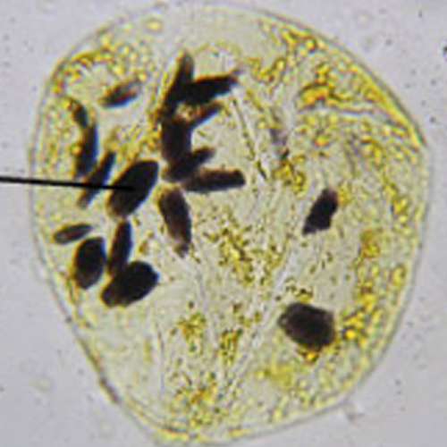
These cells contain starch grains that are stained by the common laboratory chemical – Lugol solution. Lugol solution is a solution of iodine and potassium iodide in water and it is used as an indicator test for the presence of starches, with which it reacts by turning a dark-blue/black. This solution, first made in 1829, is named after the French physician J.G.A. Lugol and is often used as an antiseptic and disinfectant.
Starches include the plant starches amylose and amylopectin and glycogen in animal cells. Lugol’s solution will not detect simple sugars such as glucose or fructose; that’s why unripe bananas are best for visualisation of starch granules.
Lugol solution is not hazardous, however, contact with skin and eyes should be avoided and the solution should not be ingested.